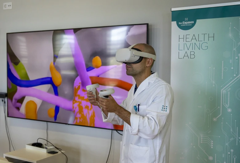
The Colorectal Surgery Unit at the Son Espases University Hospital has led a pioneering study to improve surgery for advanced rectal cancer. The work, headed by Dr Álvaro García-Granero and published in Annals of Surgery Open, introduces a digital tool that transforms MRI scans into 3D models. These models show in great detail the position of the tumour and the structures it may be invading. The precision achieved allows surgeons to plan safer approaches and preserve organs that were previously removed as a precaution.
The study included 37 patients. After reviewing the 3D reconstruction, surgeons changed their surgical plan in 38% of cases. In 24% of patients, a highly aggressive operation (pelvic exenteration) was avoided, as the model confirmed that the tumour did not affect nearby organs, even though the MRI suggested otherwise. Complete tumour removal (R0 resection) was achieved in 76% of procedures. There were no postoperative deaths and severe complications reached 27%, a very positive figure for such a complex surgery.
The project has been made possible thanks to collaboration with Cella Medical Solutions, the developer of the software used, and with the 3D Simulation and Reconstruction Unit at Son Espases.
The team is already working on further improvements. Their goal is to integrate artificial intelligence and expand this technology to other tumours, moving towards more precise and less invasive surgery.


Leave A Comment